Classification criteria and categories according to the embryo quality
Embryos obtained during in vitro fertilization (IVF) treatment are cultured in the laboratory under controlled conditions for several days. Once developed, the best quality embryo or embryos are selected to be transferred to the future mother's uterus and if there are, some are frozen for future cycles.
The success rates of an IVF process in assisted reproduction depend to a great extent on the quality of the embryo or embryos to be transferred. Those embryos of higher quality will be able to implant better in the uterus and, to do so, result in a pregnancy. Therefore, embryos are classified according to morphological characteristics such as number of cells, size, shape, etc.
This article explains the general criteria used in assisted reproduction centres to determine the quality of embryos, as well as their grading.
The different sections of this article have been assembled into the following table of contents.
Contents
- 1.
- 1.1.
- 1.2.
- 1.3.
- 1.4.
- 2.
- 2.1.
- 2.2.
- 2.3.
- 2.4.
- 2.5.
- 2.6.
- 3.
- 3.1.
- 3.2.
- 3.3.
- 3.4.
- 4.
- 5.
- 5.1.
- 5.2.
- 5.3.
- 5.4.
- 6.
- 7.
Evaluation of embryonic development
When egg fertilization with sperm is carried out in the laboratory (IVF or ICSI), the subsequent evaluation of the embryos that have been formed is fundamental.
The evaluation of the embryos quality is carried out on the basis of their morphology, that is, taking into account their shape, appearance and evolution. There are two options:
- Observation of the embryos under an optical microscope. This is the most traditional method and should be done quickly so that the embryos are out of the incubator for as short a time as possible.
- Observation of embryos using technology time-lapse. It is an incubator that records the evolution of the embryo 24 hours a day, so that the embryologist does not miss any detail of its development. This option allows the embryos to be valued without having to remove them from the incubator.
The embryos classification will allow the transfer of better quality embryos favoring the chances of achieving pregnancy. However, it is necessary to distinguish the different embryonic stages, as well as to know the appropriate nomenclature of each stage beforehand. These are detailed below.
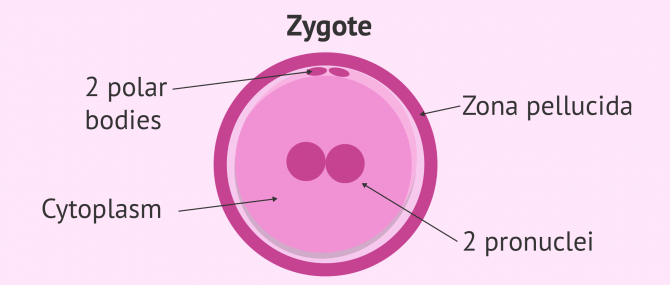
Zygote
When an IVF process is carried out, the fertilisation should be evaluated after 16-22 hours. No more time should elapse because the embryo will continue to develop. If the result is positive (fertilization has taken place) a zygote or zygocyte is observed, i.e. a fertilised ovum.
In order to assess whether zygote formation has taken place, three fundamental aspects must be observed:
- Presence of two pronuclei. These are the nuclei of the gametes (egg and sperm).
- Presence of two polar bodies containing the excess genetic material of the oocyte. These structures indicate that the meiosis of the egg has been completed.
- Uniform, clear cytoplasm.
The cytoplasm is the part of the cell that surrounds the nucleus, where the genetic material is located.
If these characteristics do not appear, the embryo is abnormal and is not considered suitable for transfer. Therefore, if 1 or 3 pronuclei and/or polar bodies are observed, the embryo should be discarded due to inadequate genetic endowment.

Embryo
When the fusion of the two pronuclei takes place, the already considered embryo initiates successive divisions.
On day 2 of embryonic development the first two divisions have occurred and the embryo has 4 cells or blastomeres. If less than 4 or more cells are observed, this is indicative of a delay or acceleration of development respectively. The evaluation of the embryo with 4 cells should be performed 44-45 hours after fertilization.
On day 3 of embryonic development the first two divisions have occurred and the embryo has 8 cells. These embryos can be transferred to the mother's uterus or continue their development in the incubator until day 5 or 6.

Morula
The morula can be observed between 90-94 hours after fertilization, which corresponds to day 4 of development. This is a conglomerate of between 12 and 16 cells reminiscent of the appearance of a blackberry.
This stage provides little information about the state of the embryo due to the compaction of all the cells. However, the degree of compaction will be important, as partial compaction results in a poor prognosis for the embryo.

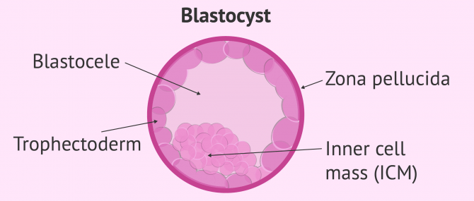
Blastocyst
The blastocyst refers to the embryo between day 5 and 6 of development. It is the last stage of embryonic development that can be carried out in the laboratory.
The blastocyst-stage embryo has about 200 cells that are gradually being restructured around the cavity that forms in the interior called the blastocele. The formation of the blastocele causes the appearance of 2 structures:
- Inner cell mass (ICM)
- responsible for originating the embryonic layers that give rise to the baby's organs.
- Trophectoderm
- It is responsible for originating the placenta.
In addition, the blastocyst has the thinnest zona pellucida. This property is related to a better exit from the zona pellucida for implantation.
Morphological criteria
Over the years, attempts have been made to unify the criteria for assessing embryo quality. For this reason, the Association for the Study of Reproductive Biology (Asociación para el Estudio de la Biología de la Reproducción -ASEBIR) has created the morphological reference criteria for assessing embryo quality.
Cell number, cell symmetry and cleavage speed
The number of cells that form the embryo is one of the most important parameters for classifying it according to its quality. It should increase from 4 cells on day 2 of development to 8 cells on day 3, etc. In this way it is said that the cleavage speed of the embryonic cells must be that which doubles the number of cells in 24 hours.
In addition, if the cells are the same size and symmetrical it will be indicative of good embryonic quality. When it comes to embryos that present cells of different sizes and with an asymmetry greater than 20% is considered non-viable embryos.
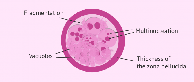
Percentage of fragmentation
It consists of small remains of cells that are arranged around the embryo making it difficult to divide and evolve.
Embryos will have a higher quality when the percentage of fragmentation is lower. Therefore, embryos without fragments or with a fragmentation percentage lower than 10% will be the first candidates to be transferred to the uterus, while if there is fragmentation higher than 35%, they are non-viable embryos.
Presence of vacuoles
The presence of vacuoles in the cytoplasm is a criterion to be taken into account when assessing embryo quality, as it is related to the degeneration and lysis of the embryo.
Vacuoles are a kind of bags that contain liquid in their interior. If they appear in the embryo in large numbers and with a large size, they can negatively influence the embryo quality.
During embryonic development, fewer vacuoles are observed on day 2 and day 3. However, there is an increase in their number from day 4 of embryonic development. In addition, most of these vacuoles are located in the trophectoderm of the embryo on day 5 .
Shape and thickness of the zona pellucida
The zona pellucida of the embryo is a round layer not too thick or thin that surrounds the embryo. It is present until the embryo reaches the blastocyst stage.
If the zona pellucida is too thick or presents anomalies, it is related to low implantation rates, as it makes it difficult for the embryo to leave. In these cases, assisted hatching should be used to facilitate the output of the internal cell mass.
Multinucleation
Each of the cells that make up the embryo must have a single nucleus where its genetic information is found. There are several reasons why human embryos contain multinucleated cells:
- Division of the nucleus not accompanied by cell division.
- Fragmentation of the nucleus.
- Abnormal migration of chromosomes in cell division, particularly during anaphase.
If cells with several nuclei appear on day 2 or 3 of embryonic development, it correlates with a lower embryo implantation rate. However, numerous studies indicate that multinucleated blastomeres only on day 3 of development do not affect embryo implantation as much.
Degree of compaction
Compaction is the process by which embryonic cells form tight joints between themselves to form the morula. It usually occurs on day 4 of embryonic development, but it is also possible on day 3.
Depending on the degree of compaction, the contour of all the cells forming the embryo may or may not be differentiated. In addition, at this time the cells cease to be totipotent and begin to specialize.
Totipotent cells are those cells capable of originating a complete organism. This is the case of the zygote or the cells formed in the first divisions of the zygote.
Embryo quality grading
Currently the embryos on day 3 and day 5 (blastocyst) of embryonic development can be classified into categories taking into account the criteria established by ASEBIR. In addition, the classification of embryos is different depending on the embryonic stage being assessed.
The selection of the best embryo for the transfer will be a determining factor in the probability of successful treatment, both in IVF treatments with own eggs and with egg donation.
Generally, the the embryos quality is indicated on the basis of four letters that are from higher to lower quality: A, B, C and D.

Grade A
Grade A embryos are considered to be of excellent quality, so the possibility of implantation is maximal. In order for an embryo to be included in this category, the parameters assessed are the following:
- 4 cells on day 2 and 7-8 cells on day 3 of the embryo.
- Blastomeres of similar size.
- Percentage of fragmentation less than 10% or null.
- Absence of multinucleated cells.
- Clear cytoplasm without vacuoles.
- Normal thickness zona pellucida.
Grade B
The grade B of embryos refers to those that have good quality with a high implantation rate. The morphological characteristics are as follows:
- 4 or 5 cells on day 2 and 7-10 cells on day 3 of the embryo.
- Slightly different sized blastomeres.
- Percentage of fragmentation around 10-25%.
- Absence of multinucleated cells.
- Clear cytoplasm with small vacuoles.
- Abnormally thick zona pellucida.
Grade C
The embryo is of intermediate quality as is the possibility of implantation. The morphological aspects that appear in this type of embryos are:
- 2 or 6 cells on day 2 and 6-12 cells on day 3 of the embryo.
- Asymmetrical blastomers.
- Percentage of fragmentation around 25-35%.
- Presence of 1-2 multinucleated cells.
- Rough cytoplasm with large vacuoles.
- Abnormally thick zona pellucida.
Grade D
Grade D embryos are of poor quality, so the likelihood of implantation is low. Their morphological characteristics are shown below:
- 3, 6 or more cells on day 2 and 3-5 cells on day 3 of the embryo.
- Asymmetrical blastomers.
- Fragmentation percentage higher than 35%.
- Crowd of multinucleated cells.
- Rough cytoplasm with large vacuoles.
- Abnormally thick zona pellucida.
Day 5 and 6 embryo classification
At this stage of development, the embryo (blastocyst) presents the inner cell mass (ICM), the cells that form the trophectoderm (TE) and the central liquid cavity (blastocele).
Three criteria are combined to classify blastocysts:
- Degree of expansion of the blastocyst. It is represented by a number from 1 to 5, 1 being an early blastocyst with null expansion and 5 being a hatched blastocyst.
- Quality of the inner cell mass indicated by a letter A-D, starting with the best quality.
- Quality of the trophectoderm with the letters A, B, C, D. Grade D indicates that the trophectoderm has cells with signs of degeneration.
FAQs from users
Can embryos survive freezing?
Currently, frozen embryos have high survival rates. However, not all embryos can be considered for freezing, as they must meet a number of requirements.
However, although there is a small risk of loss of embryos during a vitrification process, the overall results are very similar to those obtained with fresh embryos.
Can pregnancy be achieved with D-quality embryos?
Yes, pregnancy can be achieved with embryos of quality D. However, the possibilities are too low.
The Association for the Study of Reproductive Biology (ASEBIR) establishes morphological criteria for the classification of embryos according to their quality. Grade D embryos are considered to be of poor quality and have low, but not zero, implantation rates.
Whenever possible, quality A and B embryos should be transferred before grade C or D embryos.
Is embryo quality related to the health of the baby?
No. Embryo quality does not affect the health of the baby, but it is related to the possibility that the embryo implants in the uterus of the future mother and leads to a pregnancy.
Embryo quality is based on the study of morphological criteria, so it is not related to the genetic or metabolic quality of the embryo.
What is the reason for poor embryo quality?
It is quite difficult to define the exact causes of poor embryonic quality in women with good reproductive health. However, there are factors such as advanced maternal age, endometriosis, obesity, severe male factor, etc. that seem to influence the quality of embryos.
Suggested for you
In this article we have talked about embryonic development. You can continue reading here If you wish to obtain more information: In vitro embryo culture and development
We also recommend you to visit the article What Is IVF Surrogacy? – Process, Success Rates & Cost to learn more about this process.
We make a great effort to provide you with the highest quality information.
🙏 Please share this article if you liked it. 💜💜 You help us continue!
References
ASEBIR. Criterios de Valoración Mofológicos de Oocitos, Embriones tempranos y Blastocitos Humanos. 2007
Desch L, Bruno C, Luu M, Barberet J, Choux C, Lamotte M, Schmutz E, Sagot P, Fauque P. Embryo multinucleation at the two-cell stage is an independent predictor of intracytoplasmic sperm injection outcomes. Fertil Steril. 2017 Jan;107(1):97-103.e4. doi: 10.1016/j.fertnstert.2016.09.022.
Figueira Rde C, Aoki T, Borges Junior E. [Limitations and controversies in determining the predictive value of oocyte and embryo morphology criteria]. Rev Bras Ginecol Obstet. 2015 Nov;37(11):533-46. doi: 10.1590/SO100-720320150005330.
Martínez-Granados L, Serrano M, González-Utor A, Ortíz N, Badajoz V, Olaya E, Prados N, Boada M, Castilla J. Inter-laboratory agreement on embryo classification and clinical decision: Conventional morphological assessment vs. time lapse. PLoS One. 2017 Aug 25;12(8):e0183328. doi: 10.1371/journal.pone.0183328.
Salumets A, Hydén-Granskog C, Mäkinen S, Suikkari AM, Tiitinen A, Tuuri T. Early cleavage predicts the viability of human embryos in elective single embryo transfer procedures. Hum Reprod. 2003 Apr;18(4):821-5.
Yang L, Cai S, Zhang S, Kong X, Gu Y, Lu C, Dai J, Gong F, Lu G, Lin G. Single embryo transfer by Day 3 time-lapse selection versus Day 5 conventional morphological selection: a randomized, open-label, non-inferiority trial. Hum Reprod. 2018 May 1;33(5):869-876. doi: 10.1093/humrep/dey047.